


如何使用科学相机进行荧光分析
Introduction
In recent years, fluorescence analysis has become an essential technique in various scientific fields. To accurately capture and analyze fluorescent signals, scientists rely on specialized instruments such as scientific cameras. This article aims to provide insights into effectively using scientific cameras for fluorescence analysis.
Choosing the Right Camera
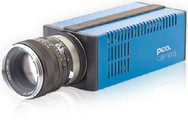
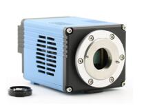
Selecting the appropriate scientific camera is crucial for fluorescence analysis. Factors to consider include resolution, noise level, sensitivity, dynamic range, and camera speed. High resolution ensures the clarity of captured images, while low noise levels allow for accurate signal detection. Sensitivity determines the camera’s ability to detect faint fluorescence, while a wide dynamic range enables capturing high-intensity signals without losing details. Lastly, a fast camera helps study dynamic fluorophore behavior accurately.
Camera Set-Up and Calibration
Proper camera set-up and calibration are vital steps in fluorescence analysis. To minimize noise and ensure optimal performance, it is necessary to adjust the settings according to experiment requirements. The exposure time should be adjusted to capture sufficient fluorescence signals without saturating the camera. Furthermore, it is essential to use fluorescence standards for calibration, allowing precise quantification of emitted light. Regular maintenance and calibration ensure accurate data interpretation.
Acquisition Software and Image Processing
Choosing appropriate acquisition software and understanding essential image processing techniques are integral for fluorescence analysis. The software should offer various features, including real-time imaging, region of interest (ROI) selection, and camera control. Additionally, understanding image processing techniques such as background subtraction, noise reduction, and contrast enhancement can significantly improve image quality and enhance data analysis capabilities.
Photobleaching and Phototoxicity Considerations
Photobleaching—loss of fluorescence due to continuous or intense illumination—and phototoxicity—caused by prolonged exposure to light—must be considered during fluorescence analysis. Adjusting camera settings, such as reducing exposure time or using low-light imaging techniques like SPIM or TIRF, can minimize photobleaching. Phototoxicity can be reduced by avoiding high-intensity illumination and implementing alternative live-cell imaging methods like fluorescence recovery after photobleaching (FRAP) or fluorescence resonance energy transfer (FRET).
Camera Maintenance and Care
Regular camera maintenance is essential for ensuring optimal performance and longevity. Keeping the camera and associated equipment clean prevents dust or debris from interfering with image quality. Additionally, regular calibration and updating camera firmware are crucial to incorporate any performance enhancements. Lastly, following manufacturer guidelines for storage and handling prolongs the camera’s lifespan.
Conclusion
In conclusion, effectively using scientific cameras for fluorescence analysis requires careful consideration of various factors. Choosing the right camera, performing proper set-up and calibration, utilizing appropriate acquisition software, understanding image processing techniques, and addressing photobleaching and phototoxicity concerns contribute to successful fluorescence analysis. Regular camera maintenance and care are vital for ensuring consistent and reliable performance. By following these guidelines, scientists can harness the full potential of scientific cameras for fluorescence analysis, enabling further advancements in various scientific disciplines.